Chemical Imaging with a
Raman Atomic-Force Microscope
PRINCIPLE OF OPERATION
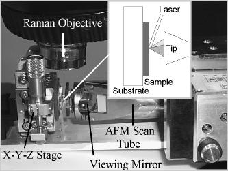
The development of a Raman-AFM depends on harnessing the complex interaction between the incident radiation, the AFM tip and the underlying sample surface. In order to produce a maximally enhanced, spatially resolved spectroscopy it would be ideal to have such a grain on the tip of the AFM. The figure above illustrates the concept. This would exploit electric field enhancement, plasmon resonance enhancement and the so-called chemical enhancement effect. Another approach is to completely metallize the tip to produce a sharp metallic probe that provides mainly field enhancement with possible chemical enhancement.
INSTRUMENT
The RAFM system is the
integration of a commercial AFM (Digital Instruments D3000) and Raman systems
(Kaiser Holoprobe) attached to an optical microscope with a modified microscope
stage to accommodate the AFM head. The laser excitation wavelength is 785 nm.
This arrangement, shown above, allows the AFM to scan a sample while side
illuminated with the Raman microprobe beam. The Raman beam size is
approximately 2-4 microns and intensity is approximately 10 mW depending on
focusing. The angle of incidence of the beam relative to the surface is grazing
at approximately 3° and can be adjusted to accommodate other angles.
The SERS active AFM tips for this study are prepared by plasma sputtering silver or gold on the conventional silicon AFM tips. This is a stochastic process and yields only a fraction ot tips with the proper alignment of metal grains to provide localized SERS. AFM analysis of a silicon test surface coated under the same conditions as the silicon tips reveals a silver grain structure averaging 40 nm in size.
RESULTS
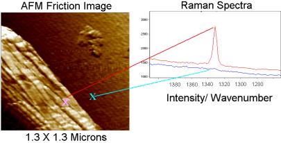
The ability of this instrument to provide spatially localized SERS was tested with diamond particles embedded in a glass microscope slide. The scanning was in contact mode for the local spectroscopic measurements. In contact mode the lateral (friction) force measurements could be simultaneously acquired along with the topography. The friction contrast was useful when measuring the diamond glass interface because the topography alone could not necessarily discern the edge. The Raman instrument was typically set for a 20-s integration time with the cosmic ray filter turned off. The 1332 cm-1 diamond peak was monitored.
The image above shows the AFM friction image of the edge of a diamond particle and the locations of the targeted SERS spectra. The diamond signal is clearly enhanced on the particle. This was observed when the tip was positioned at various orientations around the perimeter of the diamond particle. The figure below shows the signal as the AFM probe is translated across a diamond-glass boundary. In this case the background signal from the conventional Raman scattering is larger than the enhanced signal. The enhanced signal is ~15% of the total signal in this case. It is possible to find a location where the background signal is smaller. However, a typical analysis does have some background component. The spatial resolution of the spectral discrimination is estimated to be better than 50 nm. The resolution is further analyzed below. The SERS-active tips had a limited life and this was evident after scanning large, rough areas, the tips failed to produce any measurable enhancement. It is important to maintain laser focus on the tip-sample interface; any drift is evident in a steady change in the Raman signal from the silicon tip at 520 cm-1; the silicon in the tip provides an internal standard.
DISCUSSION
We are not using an extremely SERS active dye, such as rhodamine-6G. Diamond was chosen for its hardness and its distinct Raman line, not its SERS activity. The diamond on glass sample was selected because it provided a discrete edge. However, to prevent the diamond from moving it was embedded in the glass. This may have made the glass-diamond transition less abrupt and limited the apparent resolution. An accurate measurement of the resolution of the spectral discrimination requires a sample with a very discrete edge. The results presented here show approximately 50 nm resolution or better for targeting a specific region. Of course, when used in conjunction with friction or lateral force imaging of the AFM, the resolution can be an order of magnitude better at sub-nanometer resolution; the high spatial resolution (50-100nm) of targeted SERS may provide key information for interpreting the sub-nanometer chemical contrast of friction-mode AFM.
In this work we produced the tips
using a stochastic process that yield ~5-10% of the tips with the right
qualities. Silicon is probably not the best tip material due to its optical
absorption at the Raman excitation wavelength of 785nm. Apparently, this is
overcome somewhat by a relatively thick metal coating that may isolate the
SERS-active metal grain from the silicon substrate. However, the literature
indicates that the most active SERS substrates are transparent dielectrics with
relatively isolated silver grains or clusters Therefore improvements are expected if transparent (to the
excitation wavelength) dielectric AFM tips are used. Other important
optimization parameters include adjusting the tip grain size, aspect ratio and
the angle of laser incidence. A further
experimental improvement would be to use oscillation of the tip and thereby
remove conventional Raman signal from adjacent illuminated regions. Because of
the long integration times required by the CCD detector, low frequency tapping
of the surface would be required.
CONCLUSION
This combined Raman-AFM instrument has the ability to simultaneously provide high-resolution topography, friction contrast and phase contrast while scanning inside the laser beam of a Raman microprobe. We believe this combined information alone has considerable analytical utility. The further ability of the AFM tip to target the SERS effect is a significant development in analytical microscopy and has the potential for targeted single molecule spectroscopy. The unique side illumination provides the flexibility to probe opaque samples.